Super-hydrophobic surfaces are ubiquitous in nature: if you just walk in a park with ornamental ponds, you may see Lotus’ leaves floating on the water surface.

Figure 1. Droplets of water on the surface of a Lotus’s leaf. Source: Wikipedia
These aquatic plants keep their surfaces clean due to peculiar water repelling properties. A dirt free leaf surface is important for them to keep high the light-harvesting efficiency. But how the self-cleaning mechanism work? The key to this process relies on microscopic protrusions on the surface of the leaf that generate super-hydrophobicity. Before explaining the trick we need to spend some words about the behavior of liquid drops on surfaces. Water droplets on a surface of a material can spread or retain a compact almost spherical profile. The nature of the final distribution is related to the nature of the liquid (in this case water) and the surface. Surfaces can be with respect to liquid water either hydrophilic or hydrophobic. These two words are a combination of Greek words, hydros (water), -phobos (afraid), and -philos (loving), meaning water repelling and water-loving tendency, respectively. Contrary to a hydrophilic one, a hydrophobic surface does not allow a small drop of water to easily spread on it.
If you try to place a drop of water on microscope glass slide and align its side to your eye level, you will see the droplet profile on the surface has the profile of an almost spherical cap. The contact of the droplet with the surface is characterized by an angle between the water surface and the surface. This angle is called Young angle in honor of the English scientist Thomas Young (1773-1829) that first studied systematically the phenomenon of wetting. This angle is the results of the surface energy of the different materials involved in the contact: air, water, and glass. When the force acting on the different materials are balanced then the contact angle takes an equilibrium value. In the following box, you can try to measure the approximate value of the contact angle of a liquid on a surface using a smartphone camera and a computer software.
MEASURING THE CONTACT ANGLE OF A LIQUID DROPLETThe contact angle can be approximately determined by taking a picture of the droplet on the surface and by measuring the angle with software for image analysis. A smartphone camera can be a good starting if supplied with a small magnifier lens and good lighting. The figure below shows the image of a droplet of water on the surface of a microscope glass slide. The photo was taken with a smartphone camera aligned to the side of the slide. The lighting can be improved to make sharper the border of the photo. 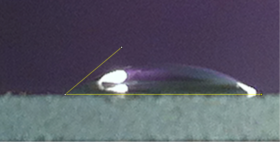 Figure IB: Droplet of water on a glass surface.The contact angle can be measured from the photo using the public domain software ImageJ. This is a powerful program developed at NIH in the USA that can be used to perform sophisticated image analysis. One popular distribution is called Fiji (available at imageJ.net) and it contains a customized version of ImageJ with a collection of selected plugins.  Once the image is load by the program, it is possible to use the angle tool to calculate the contact angle using the angle tool by clicking the 6th icon in the control bar of the program.  The angle is given by the baseline and the approximate tangent to the contact point of the drop with the surface, as shown in the Figure IB. |
The cosine of the angle is given by the value of these forces by the aforementioned Young equation:

The 

Figure 2. Two extreme cases of contact angles. On the left, a water drop on a superhydrophobic surface, on the right on a hydrophobic surface.
The wetting of flat surfaces is controlled by the value of 
On the lotus’ leaf surface, the contact angles are much larger than 90o producing what is called a superhydrophobic surface. This effect is a consequence of the peculiar microscopic morphology of the leaf surface. If you give a closer look with an electron microscope to the lotus’ leaf surface (Figure 3) you will be surprised to see that the surface is far being flat.

Figure 3. The magnified surface of a rose petal. Source: Wikipedia.
You can see a myriad of small protruding pegs like the nail bed of an Indian fakir. The roughness of the surface traps in different extend air bubble that alter the properties of the surface. Indeed, to mathematically describe the wetting properties, we need to use slightly different equations for the contact angle. This trick has been used in several interesting variations by a plethora of different species of plants and animals (see [1] for a recent review). We are now trying to learn from nature to exploit the same principle for nanotechnological applications. The patterning of the surface with microscopic protrusions has been used to design bio-inspired super-hydrophobic material as for example, in the form of silica pillars as in the following graphics representation.

Figure 4: Superhydrophobic surface obtained using silica micropillars.
How small protrusions or holes or molecular surfaces can be still retaining the superhydrophobic effect? In order to theoretical answer this question, molecular dynamics simulations studies of water near structured hydrophobic surfaces have been performed [2]. The surface of a crystal of n-eicosane molecules was used as a model of the hydrophobic surface. The planar crystal surface was used as a reference. The structured alkane surfaces have been built by modeling a hole and a protrusion of approximately 2.5 nm diameter and 0.5 nm depth or height, respectively. Layers of water molecules have been used to cover both crystal surfaces and MD simulation of several nanoseconds (see Figure 6) have been used to study the wetting behavior. Different calculated properties have shown that the insertion of surface structuring features increases their hydrophobicity. In particular, the water density is reduced near both the structural features, resulting in a decrease of the number of residual contacts between water and the surface of about 40% with respect to the planar surface. In addition, the calculated interfacial energy of the structured surfaces is about $7 mJ m^2$ higher than the planar surface. All these results indicate that the hydrophobicity relative to the planar surface increases by a similar amount for both structured ones [2].

Figure 5. Simulation of water on the pitted polymeric surface [2]. Courtesy of Dr. Sandeep Pal.
Oct 2022. UPDATE: AN INSTRUCTABLE ON THIS TOPICS IS ALSO AVAILABLE HERE.
REFERENCE
- T. Darmanin and F. Guittard. Superhydrophobic and superoleophobic properties in nature. Material Today. Vol 18 (5), 273, (2017).
- S. Pal, D. Roccatano, H. Weiss, H. Keller, F. Müller-Plathe. Molecular dynamics simulation of water near structured hydrophobic surfaces: interfacial energies. ChemPhysChem, 6, 1641-1649, (2005).


Pingback: EXPLORING THE LOTUS EFFECT USING CANDLE SOOT