Silica is an essential component of many everyday materials. We use silica in containers, appliances, and electronic devices such as smartphones, computers, and tablet screens. These surfaces come into frequent contact with our skin, providing ample opportunities for pathogens, viruses, and microbes to accumulate. Therefore, it is crucial to study the adsorption mechanisms of these microorganisms on silica surfaces.
In collaboration with experimental groups at the University of Lincoln, the University of Greenwich, and Diamond Light Source, we investigate the adsorption mechanisms of isolated RBD regions of the COVID-19 spikes on silica nanoparticles. Our study proposes a simple geometrical model for packing protein particles on spherical silica nanoparticles, which aligns well with the available experimental data. This model revealed a surface occupancy of 32% relative to the maximum theoretical RBD packing, indicating significant adsorption.
My computational contribution to this study involved using molecular dynamics (MD) simulations to explore the binding modes and orientations of the protein adsorbed on a model silica surface. The model consisted of a flat patch of silica surface, which serves as a good approximation of the nanoparticle surface used in the experimental part of this study, given their size. Our findings showed that up to 25% of the RBD’s secondary structures underwent conformational changes as a result of adsorption onto silica nanoparticles.
These insights enhance our understanding of the principles governing protein-surface interactions and can contribute to strategies for controlling the spread of SARS-CoV-2 through contaminated objects.
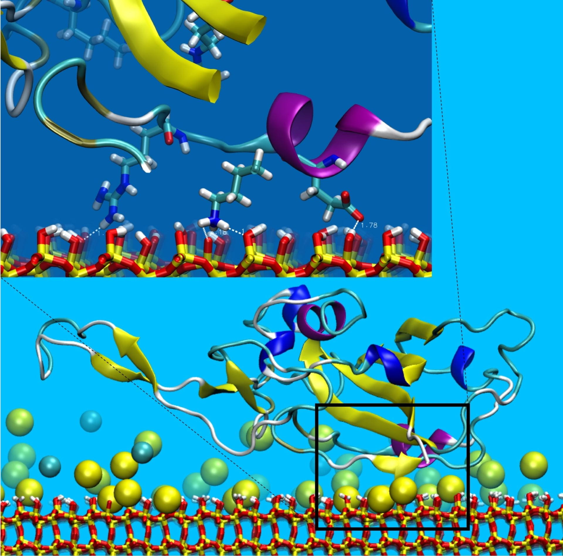
MD simulations of RBD-silica interaction. (A) Representation of the RBD adsorbed on silica in a representative configuration at the end point of simulation 1. The rectangle highlights the area of contact detailed in panel B. (B) Detailed representation of three residues in close contact with silica: arginine 346 (R346), lysine 356 (K356) and glutamic acid 340 (E340) from left to right. The distances
between hydrogen and oxygen atoms at the interface are indicated and are compatible with the hydrogen bonds length.

